


Electrocardiogram (ECG)
A diagnostic test used to record the electrical activity of the heart
Some heart diseases can lead to a disturbance in the electrical activity of the heart, prompting doctors to perform an electrocardiogram (ECG) to detect and diagnose them, especially when specific symptoms are present, such as chest pain, difficulty breathing, palpitations, or irregular heartbeats. The ECG is one of the necessary tests that detect health problems related to the electrical function of the heart, and it is one of the simple and painless tests that record the electrical activity of the heart. The heartbeats are generated from electrical impulses produced in the upper part of the heart, then transmitted to the lower part. This electrical activity is detected by attaching several electrical electrodes to different skin areas to record the heart’s electrical activity.
As is known, the heart can convert electrical work from electrical signals to a graph drawn on paper by recording heart rhythms, and the electrical activity makes the heart contract. Therefore, it is possible to identify problems that your heart may be exposed to regarding rate and rhythm when measuring it.The heart has an electrical system that helps it contract and relax regularly and pump blood. A specialized cardiologist can analyze the results of an electrocardiogram immediately during the test, which takes no more than five minutes and is painless for the patient.
Doctors rely on electrocardiograms to diagnose several conditions, including:
- Examining the electrical function of the heart
- Checking the heart’s safety in the presence of risk factors for heart disease, such as chronic diseases like diabetes, high blood pressure, high cholesterol, smoking, or a family history of heart disease.
- Diagnosing heart rhythm disorders
- Diagnosing the cause of chest pain, which may be due to acute heart failure, pericarditis, myocarditis, or angina pectoris
- Diagnosing the causes of symptoms related to heart disease, such as chest pain, shortness of breath, palpitations, and dizziness
- Diagnosing complications of heart disease, such as heart enlargement, heart failure, and heart rhythm disorders.
Electrical System of the Heart
Each heartbeat of the heart starts with an electrical signal generated by specialized cells located in the heart’s right atrium, called the sinoatrial node. This node is connected to the left atrium via an electrical pathway, which causes both atria to contract simultaneously. Then, the nerve signal travels to what is called the atrioventricular node, which slightly delays the electrical impulse before it reaches the ventricles via nerve fibers to cause their contraction. It should be noted that these electrical pulses are produced very precisely and coordinated. An electrocardiogram (ECG) test can detect any disruption in this coordination.
How is the procedure of an electrocardiogram performed?
This device can capture electrical signals and convert them into a graph. Wires from an electrocardiogram (ECG) machine are attached to electrodes, which are metal strips that conduct electricity. The electrodes are placed on each arm and leg and at six points on the chest above the heart. These electrodes pick up the electrical impulses produced by the heart with each beat and transmit them to an amplifier within the ECG machine. The amplified signals then flow through a thin wire coil suspended in a magnetic field, causing the wire to move due to the interaction between the electrical signals and the magnetic field. A sensitive lever records the movement of the wire onto a moving graph paper, resulting in a graph of the heart’s electrical activity known as leads.
Each heartbeat produces a series of waveforms; a regular heartbeat has a specific waveform pattern. Certain types of heart disease can recognizably alter this known pattern. Each electrode connected to the body is like a camera, and the doctor places ten electrodes on the human body – one on each arm, one on each leg, and at six points on the chest – thereby obtaining ten cameras or electrodes from different directions. Each camera produces another image of the heart because each one captures a different angle of the same view.
What does the electrocardiogram (ECG) reveal?
After completing the heart monitoring and reading the results, a doctor can detect many heart-related diseases. They can discover if the patient is suffering from acute heart failure, cardiac arrhythmias, enlarged heart muscle, pericarditis, myocarditis, or an imbalance in the body’s salt levels. The monitoring takes the form of waves with several sections that come out of the device, and the doctor examines them to see the progress of the electrical signal in the heart. The doctor then reads and analyzes the waves.
An electrocardiogram (ECG) can reveal disorders in the production and conduction of the electrical wave. It can also indicate conditions resulting from an imbalance in the body’s salt levels, drug toxicity, or irregular heart function. Changes in heart monitoring may reveal the presence of heart muscle failure, whether recent or old.
The electrocardiogram can examine the heart rate to determine if there is an irregularity in the heartbeat’s speed. Heart monitoring also helps determine the heart rate and rhythm to determine if it is fast or slow.
Heart monitoring can also detect evidence of a previous heart attack and the extent of damage to the heart or determine if blood and oxygen are not reaching the heart adequately. Additionally, an electrocardiogram can identify distortions in the heart’s walls, heart defects, and other heart problems.
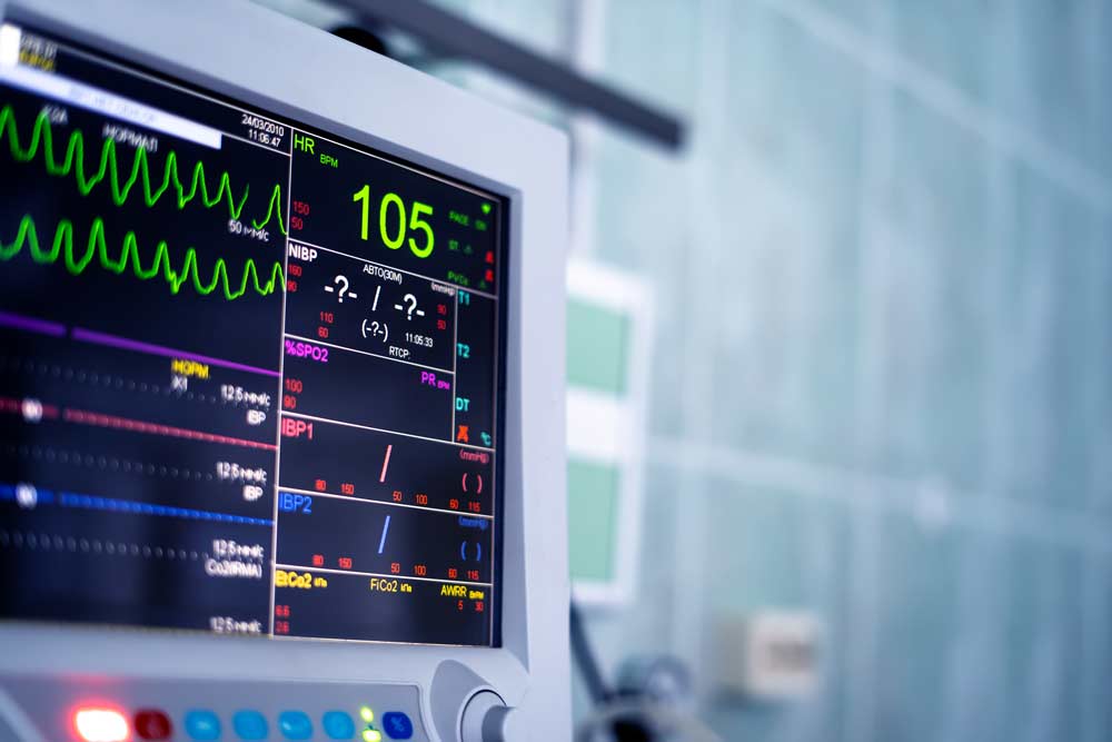
When the results are normal, the heart rhythm is at a rate of 60-100 beats per minute.
However, if the results are abnormal and indicate the presence of heart disease, the results appear as follows:
- Slowing the heart rate to less than 60 beats per minute.
- Acceleration of the heart rate to more than 100 beats per minute.
- Irregular heart rhythm.
- Specific changes in the electrocardiogram may indicate several diseases, most notably the elevation or depression of the ST segment, which in most cases means acute heart failure.
It is necessary to point out in this field that this type of examination is useless in angina cases requiring other tests for diagnosis. Additionally, in some cases, especially with a family history of heart disease, the result of an electrocardiogram alone does not indicate the presence or absence of heart disease. The patient’s medical history, physical examination, electrocardiogram, and other tests should be used to diagnose the patient’s condition. An electrocardiogram does not indicate the possibility of heart failure.
An exercise test is used to diagnose angina. During this test, the person performs physical work, which can cause symptoms to appear if there is heart disease and changes in the electrocardiogram. Many factors can affect the test’s accuracy, such as poor electrode adhesion, movement during the test, exercise before the test, anxiety, or deep breathing during the test. Some changes in the electrocardiogram may not appear until several hours after symptoms occur, so it is essential to monitor the patient’s condition and perform an electrocardiogram after several hours. In some cases, changes in the electrocardiogram may appear after physical exertion, and this test is called an exercise test.
In many cases, some changes in the electrocardiogram are permanent and known due to heart disease. Therefore, older adults and heart disease patients are advised to carry an electrocardiogram with them permanently when visiting a doctor or having an electrocardiogram. Reviewing previous electrocardiograms and comparing them to the current one is essential for these individuals.