


Peripheral Artery Disease
Advanced methods of treatment that prevent serious complications
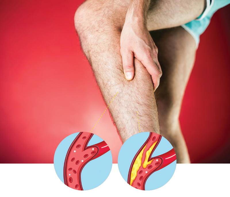
Arteries are blood vessels that carry blood rich in oxygen throughout your body. Healthy arteries have smooth inner walls and blood flows through them easily. Some people, however, develop clogged arteries, which result from a build-up of a substance called plaque on the inner walls of the arteries. Arterial plaque can reduce blood flow or, in some instances, block it altogether.
Peripheral artery disease (PAD) is a common circulatory problem in which narrowed arteries reduce blood flow to your limbs. When you develop PAD, your extremities – usually your legs – don’t receive enough blood flow to keep up with demand. If left untreated, PAD can lead to gangrene and amputation. However, modern methods of treatment have been accompanied by state-of-the-art equipment to treat the hardening or blockage of peripheral arteries; therefore, it is rare nowadays to find a patient who got gangrene which causes the amputation of the foot.
PAD is usually caused by a build-up of fatty deposits in the walls of the leg arteries. The fatty deposits, called atheroma, are made up of cholesterol and other waste substances. The build-up of atheroma on the walls of the arteries makes the arteries narrower and restricts blood flow to the legs.
Many people with PAD have no symptoms. However, some develop a painful ache in their legs when they walk, which usually disappears after a few minutes’ rest. The pain can range from mild to severe, and usually goes away after a few minutes when you rest your legs. Both legs are often affected at the same time, although the pain may be worse in one leg.
Causes
People with diabetes are at higher risk of developing atherosclerosis, the most common cause of peripheral artery disease. Factors that increase your risk of developing peripheral artery disease include smoking, diabetes, obesity, high blood pressure, high cholesterol, increasing age, a family history of peripheral artery disease and heart disease or stroke. If the blockage remains in the peripheral arteries in the legs, it can cause pain, changes in skin color, sores or ulcers and difficulty walking. Total loss of circulation to the legs and feet can cause gangrene and loss of a limb. If the blockage occurs in a carotid artery, it can cause a stroke.
Symptoms
While many people with peripheral artery disease have mild or no symptoms, some people have leg pain when walking (claudication). Claudication symptoms include muscle pain or cramping in your legs or arms that’s triggered by activity, such as walking, but disappears after a few minutes of rest. The location of the pain depends on the location of the clogged or narrowed artery. Calf pain is the most common location. The severity of claudication varies widely, from mild discomfort to debilitating pain. Severe claudication can make it hard for you to walk or do other types of physical activity.
Symptoms also include painful cramping in your hip, thigh or calf muscles after certain activities, such as walking or climbing stairs, leg numbness or weakness, coldness in your lower leg or foot, sores on your toes, feet or legs that won’t heal, a change in the color of your legs, among others. If peripheral artery disease progresses, pain may even occur when you’re at rest or when you’re lying down (ischemic rest pain). It may be intense enough to disrupt sleep. Hanging your legs over the edge of your bed or walking around your room may temporarily relieve the pain.
Peripheral angioplasty
A peripheral angiogram is a test carried out to identify any blood vessel narrowing or blocked areas in the arteries. By injecting a dye (contrast material) into your blood vessels, this test allows your doctor to view blood flow through your arteries as it happens. Your doctor is able to trace the flow of the contrast material using imaging techniques, such as X-ray imaging or procedures called magnetic resonance angiography (MRA) or computerized tomography angiography (CTA).
Catheter angiography is a more invasive procedure that involves guiding a catheter through an artery in your groin to the affected area and injecting the dye that way. Although invasive, this type of angiography allows for simultaneous diagnosis and treatment — finding the narrowed area of a blood vessel and then widening it with a dilating procedure or administering medication to improve blood flow.
Before the procedure you will be given medicine to help you relax. You will be awake, but sleepy. You may also be given blood-thinning medicine to keep a blood clot from forming and you will lie down on your back on a padded operating table. Your surgeon will inject some numbing medicine (local anesthesia) into the area that will be treated, so that you do not feel pain. Your surgeon will then place a tiny needle into the blood vessel in your groin. A tiny flexible wire will be inserted through this needle.
Your surgeon will be able to see your artery with live x-ray pictures. Dye will be injected into your body to show blood flow through your arteries. The dye will make it easier to see the blocked area.
- Your surgeon will guide a thin tube called a catheter through your artery to the blocked area.
- Next, your surgeon will pass a guide wire through the catheter to the blockage.
- The surgeon will push another catheter with a very small balloon on the end over the guide wire and into the blocked area.
- The balloon is then filled with contrast fluid to inflate the balloon. This opens the blocked vessel and restores blood flow to your heart.
- A stent may also be placed in the blocked area. The stent is inserted at the same time as the balloon catheter. It expands when the balloon is blown up. The stent is left in place to help keep the artery open. The balloon and all the wires are then removed.
The entire process takes between half an hour and an hour, but in some cases it may take longer if the doctor decides to treat the blockage of the artery by dilating it or injecting drugs to dissolve the blood clot if present.


I enjoy the article
It works really well for me