Dr. Mohamad Mansour
Head of Non-Invasive Cardiology, Clemenceau Medical Center Beirut (CMC) Introduces Vivid Pioneer Ultrasound
Next-Generation Cardiac Imaging: Greater Precision and Smarter Structural Heart Interventions
As part of its ongoing commitment to investing in cutting-edge medical technologies, Clemenceau Medical Center (CMC) Beirut has introduced the Vivid Pioneer Ultrasound, one of the world’s most advanced echocardiography systems. Combining advanced four-dimensional imaging with artificial intelligence, the system delivers exceptional image quality, improves diagnostic accuracy, supports complex structural heart interventions, and further enhances the quality of cardiovascular care at CMC. In this interview, Dr. Mohamad Mansour, Head of Non-Invasive Cardiology at CMC Beirut, discusses the clinical significance of this latest technology, its key innovations, and its impact on patient care and contemporary cardiac imaging.
What prompted CMC to introduce the Vivid Pioneer Ultrasound system?
CMC has always been committed to investing in technologies that bring tangible clinical value and enhance the quality of patient care. The introduction of the Vivid Pioneer Ultrasound reflects this philosophy, allowing us to offer one of the world’s most advanced echocardiography systems only shortly after its launch in the United States and Europe.
As one of the first medical centers in Lebanon and the Middle East to adopt this next-generation platform, CMC continues to strengthen its cardiovascular imaging capabilities while providing physicians with advanced diagnostic tools that support greater accuracy, more efficient workflows, and improved patient outcomes.
What key innovations distinguish Vivid Pioneer Ultrasound from earlier systems?
The Vivid Pioneer Ultrasound offers a substantial improvement in image quality and spatial resolution, allowing physicians to visualize cardiac anatomy and function with unprecedented clarity.
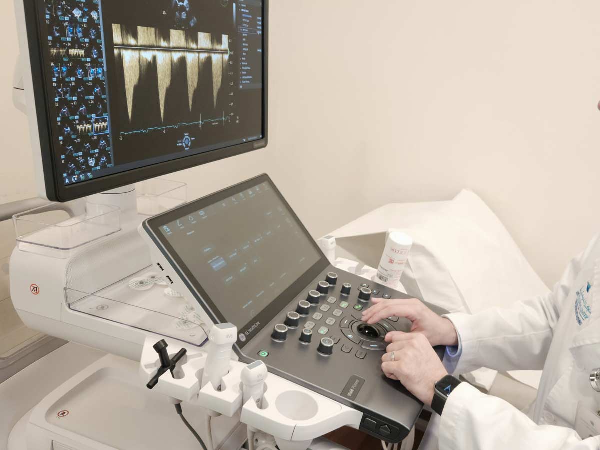
The system delivers highly detailed 4D echocardiographic imaging while incorporating AI-powered workflow tools that enable faster and more accurate quantitative measurements during examinations. These capabilities are particularly valuable during structural heart interventions, including MitraClip procedures for mitral valve repair, TriClip procedures for tricuspid valve repair, and left atrial appendage closure (LAAC), where precise imaging and accurate measurements are essential for procedural success.
In addition, the platform offers a sophisticated interventional echocardiography environment that provides real-time procedural guidance, allowing the heart team to obtain rapid and highly accurate measurements throughout the procedure while supporting more confident clinical decision-making.
What role does artificial intelligence play in the Vivid Pioneer platform?
The system incorporates an integrated AI technology supported by advanced deep-learning algorithms, significantly improving efficiency, measurement consistency, and overall diagnostic confidence.
With a single click, the platform can automatically perform many routine cardiac measurements with high accuracy, reducing operator variability while accelerating reporting and improving workflow efficiency. The software is also designed for continuous upgrades, allowing the system to benefit from future technological developments through regular software updates.
Importantly, artificial intelligence is intended to support, not replace, the physician. Clinical expertise remains the cornerstone of every examination as well as preparing the final medical report. Cardiologists continue to acquire the images, verify all measurements, interpret the findings, and make the final clinical decisions. AI serves as a powerful assistant that enhances speed, standardization, and accuracy while leaving medical judgment firmly in the hands of the clinician.
Which patients are most likely to benefit from the Vivid Pioneer Ultrasound?
Virtually any patient requiring echocardiographic assessment can benefit from the Vivid Pioneer system, whether undergoing routine cardiac evaluation or more complex diagnostic investigations.
Its greatest value, however, is seen in patients requiring highly precise quantitative assessment, particularly those being evaluated for structural heart interventions. AI-assisted 4D imaging enables highly accurate reconstruction and analysis of cardiac structures, providing physicians with comprehensive anatomical and functional information.
Can the Vivid Pioneer Ultrasound reduce the need for other cardiac imaging modalities?
The answer depends on the individual clinical scenario. For many patients, the outstanding image quality provided by the Vivid Pioneer Ultrasound supplies all the diagnostic information required, reducing the need for additional imaging studies that were previously necessary because of suboptimal echocardiographic visualization.
Nevertheless, in selected complex cases, cardiac magnetic resonance (CMR) or other advanced imaging modalities may still be indicated for further evaluation.
There is no doubt, however, that the Vivid Pioneer system represents a significant leap forward in echocardiographic imaging, enabling far more detailed assessment of myocardial function, cardiac valves, and overall cardiac function than was previously possible.
How has the Vivid Pioneer Ultrasound improved imaging in traditionally challenging patient populations?
Yes. Patients with chronic lung diseases, particularly chronic obstructive pulmonary disease (COPD), have traditionally presented challenges for echocardiographic imaging because lung hyperinflation often limits ultrasound penetration and image quality.
With the Vivid Pioneer Ultrasound, image quality has improved substantially, allowing visualization of anatomical details that were previously difficult—or sometimes impossible—to assess. While image acquisition still depends on individual patient characteristics, the improvement over previous-generation systems is considerable and increases diagnostic confidence.
How has the Vivid Pioneer Ultrasound transformed the way cardiac function is assessed?
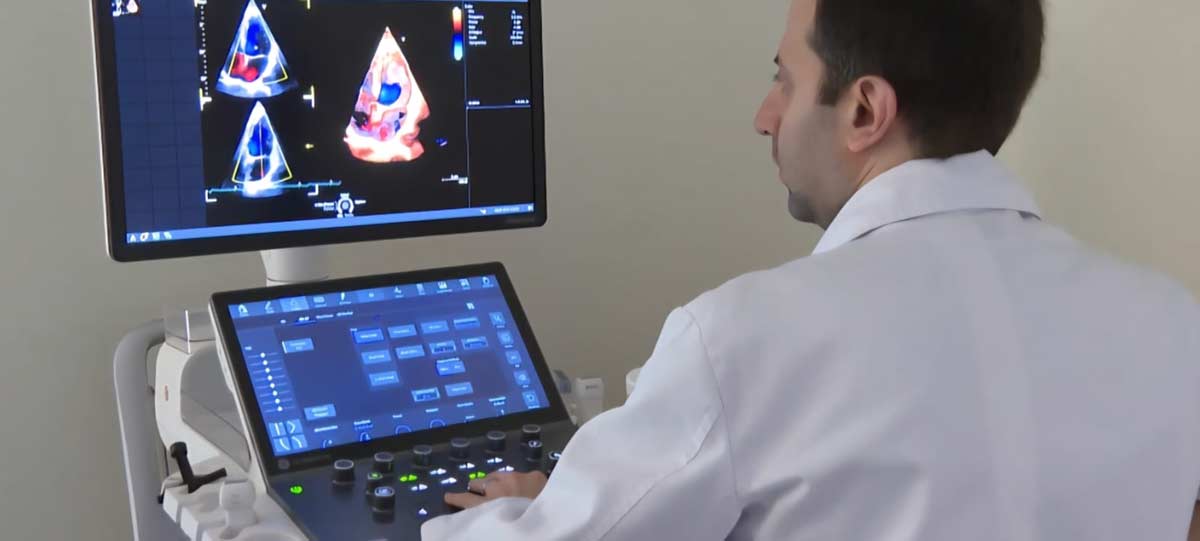
Today, technology allows us to visualize the heart with an unprecedented level of realism that was unimaginable only a few years ago.
The system provides superior visualization of the myocardium, cardiac valves, and surrounding cardiac tissues while delivering a more comprehensive assessment of cardiac function. Advanced 4D imaging enables detailed reconstruction and analysis of cardiac anatomy, giving physicians a deeper understanding of each patient’s condition and facilitating more precise therapeutic planning.
What message does CMC wish to convey through the introduction of this technology?
At Clemenceau Medical Center, investing in advanced technology is not simply about acquiring the latest equipment; it is about adopting innovations that deliver meaningful clinical value for patients.
Technology achieves its full potential only when combined with the expertise of highly experienced physicians who know how to maximize its capabilities. By integrating world-class medical expertise with the latest advances in cardiovascular imaging, CMC continues to provide more accurate diagnoses, safer procedures, and patient-centered cardiovascular care that meets the highest international standards.