


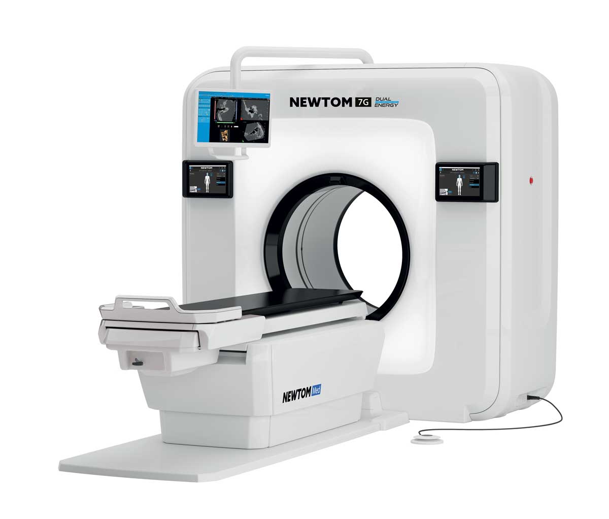
NEWTOM 7G for excellence in MSK imaging
Back in 1996, NEWTOM – once known as QR and based in Verona, Italy – released the first-ever CBCT device for clinical imaging, marking the start of three decades as a pioneer in the dental sector. Since then, the brand has extended its scope to both the medical and veterinary fields, introducing game-changing technologies along the way. One of the most recent solutions combines the NEWTOM 7G, a Multi-Scan Body CBCT device, with Dual Energy technology to elevate diagnostic performance.
CBCT technology and the benefits of Dual Energy
Thanks to its Dual Energy system, NEWTOM 7G ensures several additional benefits that support imaging professionals with their diagnostic needs. Greater precision, improved tissue contrast, and fewer artifacts are three key aspects that will underpin the excellence of the MSK imaging capabilities enabled by this superb device.
Dual Energy technology enhances diagnostic performance, and NEWTOM 7G enables physicians to use Cone Beam technology to investigate all areas of the body, including the spine, shoulder, and hip. Suited to patients with a cumbersome build, the patient table can handle up to 215 kg. Functions and automatisms are also included to adapt FOVs and X-ray doses to the patient’s build, particularly in children, for whom lower doses are recommended. With a resolution of up to 90 µm, small complex structures such as the inner ear can be analysed with the utmost precision.
Performance, ergonomics and safety in a highly versatile device
NEWTOM 7G generates ultra-HD volumetric images with native isotropic voxel resolution, non-overlapping sections and fewer artifacts. Unlike the spiral fan beam scan typical of other MSCTs, a single cone beam scan increases image quality, contains the X-ray exposure area and reduces costs. The high-power X-ray generator with rotating anode and small focal spot (0.3 mm) maximises performance and ensures emissions can always be adapted to specific needs. The large, latest-generation HD flat-panel detector with a high signal-to-noise ratio yields improved soft-tissue display. Innovative volumetric reconstruction algorithms and advanced filters minimise reconstruction times and optimise imaging.
Multiple acquisition protocols are available, ranging from static Ray2D examination to the investigation of joint dynamics with the CineX protocol and in-depth 3D volumetric diagnosis with ultra-high definition of bone tissue. Equipped with a high-power generator (120 kV – 20kW) and a high-sensitivity 3D panel, the 7G features several innovative algorithms for volumetric reconstruction. Beyond performance considerations, NEWTOM has also devoted its attention to ergonomics and the practical side of a physician’s activities, optimising examination flow thanks to the on-board 10” touchscreen control panel and the latest software suite with processing, sharing and RIS/PACS connectivity functions. The 7G safeguards patient health and comfort as they lie comfortably on the motor-driven table. Doses are always proportioned to the patient’s build and the type of examination.
Suitable for radiologists and orthopaedic, otolaryngology and dentistry specialists, NEWTOM 7G lets you capture accurate information on bone tissues – micro-structures included – to gain an in-depth understanding of the overall clinical picture, optimising analysis of treatment outcomes, at lower costs compared to conventional procedures. Exclusive automatisms optimise workflows and avoid the variability and uncertainty inherent in manual examination control, especially during patient positioning and actual exposure.
How does Dual Energy technology elevate diagnostic performance?
![]()
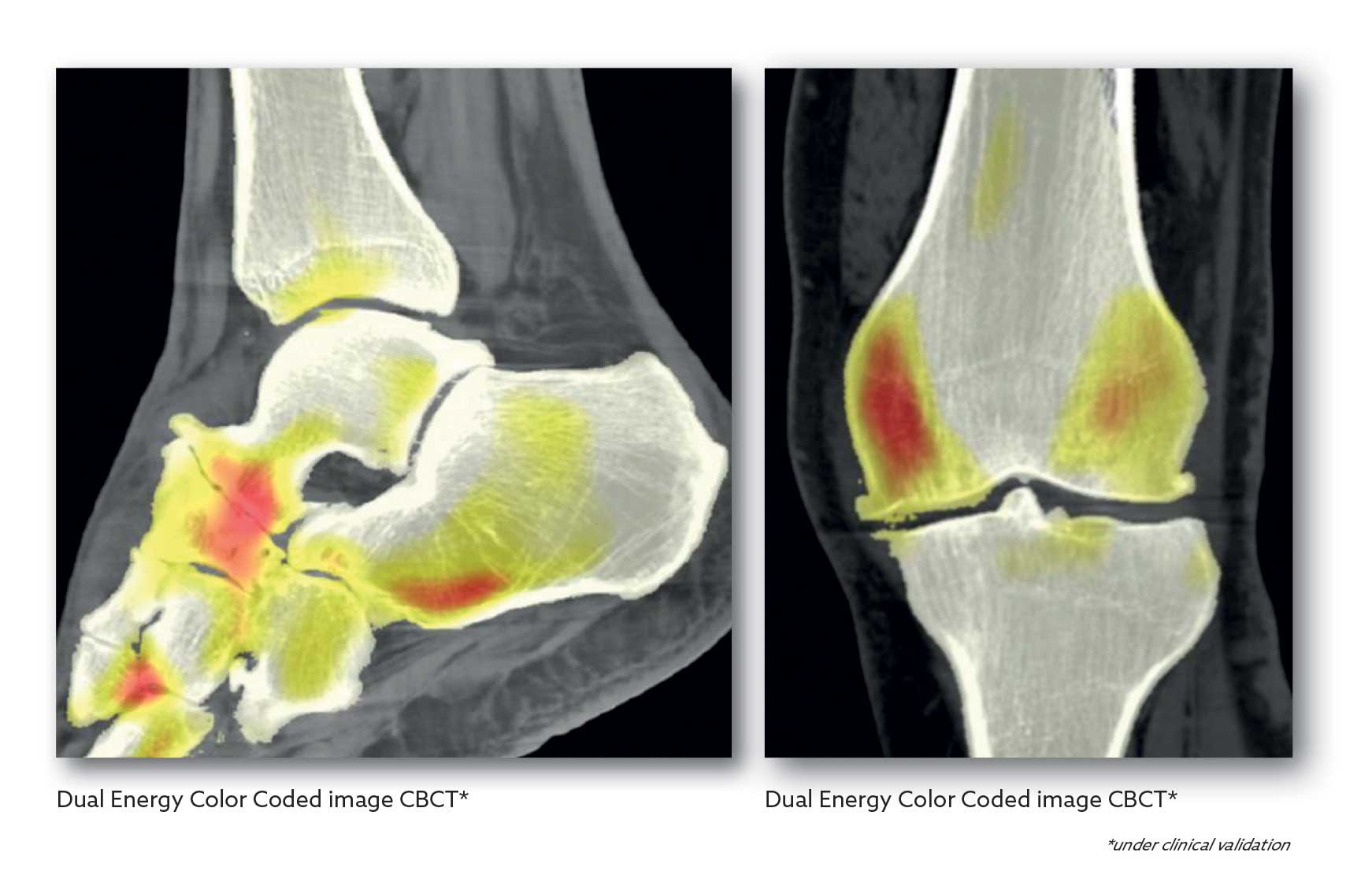
Dual Energy, as the name suggests, uses two different radiant energies that acquire two sets of images of the same anatomical area. Since tissues possess different sensitivities to different energy levels, the images that are obtained are able to return information regarding the chemical composition of the tissues themselves. This enhances the confidence and precision with which pathologies are detected.
Reconstruction of the information from the low and high energies is instantaneous. The special DE software developed by NEWTOM allows real-time selection of energy levels during display. This means you can access a vast spectrum of information, making pathology detection more effective and reliable.
This makes it ideal when you need superior quality visualisation of cortical bone, trabecular bone and provides significantly better quality soft tissue images. Dual Energy also mitigates beam-hardening artifacts, a recurring issue in tomography and one that makes it difficult to make a clear diagnosis. By being able to reconstruct virtual monochromatic images at several keV levels, Dual Energy improves soft tissue visualisation, reduces metal artifacts and provides a basis for tissue characterisation, ultimately leading to excellent image quality.
NEWTOM’s Dual Energy software enables users to perform semi-automatic tissue segmentation and the application of colour-coded images, streamlining the differentiation of materials within the scanned area. This allows rapid and straightforward detection of potential pathologies.
Quantitative tests have demonstrated that, compared to single-energy CBCT exams, Dual Energy protocols improve HU accuracy, contrast resolution and image homogeneity.
One of the causes of image quality decay is the appearance of artifacts caused by patient movement. NEWTOM has developed an algorithm that corrects any movement-induced distortions. It does this on each axis, offsetting any shifts, rotations or variations in distance from the panel. So, the new features enable weighted blending of high- and low-energy imaging. The resulting images combine the low noise typical of high-energy acquisition with the high-contrast resolution of the low-energy image set. NEWTOM’s Dual Energy technology provides unique clinical information that lets you highlight, characterise, quantify and distinguish tissues, which have different sensitivities to different energy levels, within the areas being scanned. This delivers supplementary information about the chemical composition of the materials in the area being examined, thereby helping physicians make more accurate diagnoses.
In conclusion, Dual Energy technology takes musculoskeletal imaging to a new level. It improves soft tissue differentiation and reduces artifacts. Diagnostic accuracy and patient care are major benefits when applying this technique in the field of orthopaedics. For more details or to discuss your needs with our team, complete the online form and we will contact you.